Kaiba's doctors contacted Dr. Glenn Green, who then got emergency permission from the Food and Drug Administration to do a special procedure which, before Kaiba, had only been tried on animals. Luckily for everyone involved, the surgery was a sucess!
"It's magical to me," said Dr. Glenn Green, an associate professor of pediatric otolaryngology at the University of Michigan who implanted the splint in Kaiba. "We're talking about taking dust and using it to build body parts."knew they had to come up with something, or the alternative would be a life on a ventilator.The doctors used a laser 3D printer to create a splint to hold open the baby's collapsing airway. It was a few centimeters long and 8 millimeters wide and made of polycaprolactone or PCL.
When a splint is created using PCL, it becomes a sort of biological placeholder, propping up structures while the body heals around it.Kaiba is now 20 months old and breathing on his own. (see cute pic of him here) Doctors say it will take the implant 3 years to degrade and then, presumably, the trachea will continue growing normally.
It's worth reading the whole article. Or, you can read the published paper in the New England Journal of Medicine, "Bioresorbable Airway Splint Created with a Three-Dimensional Printer"!
From Figure 1 in the publication...
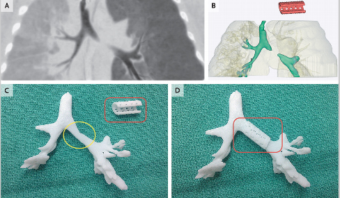
Panel A shows the airway in expiration before placement of the splint; the image was reformatted with minimum-intensity projection. Panel B shows the patient-specific computed tomography–based design of the splint (red). Panel C shows an image-based three-dimensional printed cast of the patient's airway without the splint in place, and Panel D shows the cast with the splint in place.
UPDATED 6.26.13 This University of Michigan Health Systems article has more detail on this story - well worth a read. It also includes this video:
Th article clarified for me that the splint was "sewn around Kaiba’s airway to expand the bronchus and give it a skeleton to aid proper growth. Over about three years, the splint will be reabsorbed by the body."
“The material we used is a nice choice for this. It takes about two to three years for the trachea to remodel and grow into a healthy state, and that’s about how long this material will take to dissolve into the body,” says Hollister.
This article also states that severe tracheobronchomalacia is rare: Of the 1 in 2,200 babies born with tracheomalacia, most grow out of it by age 2 or 3, although it often is misdiagnosed as asthma that doesn’t respond to treatment. Severe cases, like Kaiba’s, are about 10 percent of that number.
If you are interested, the University of Michigan Pediatric Medical Device Consortium (M-PED) created a 28-minute video entitled "Innovative Pediatric Manufacturing: Strategies & Solutions - A Guide To Bringing Your Device To Market" which discusses FDA requirements for manufacturing medical devices for children (and adults).
No comments:
Post a Comment